Pages

Wednesday, October 31, 2012

A few tech summaries from FiO


View from the convention center
Brief technical summaries of some talks that might interest folks I know:
1)      X-ray crystallography generally requires monochromatic, spatially coherent beams to determine crystal structure through diffraction patterns. It is difficult to produce such perfectly coherent beams. Emil Wolf, University of Rochester, gave a single author talk on a general formula which gives the effect of spatial degree of coherence of incident radiation on the diffraction pattern in X-ray crystallography. This enables the use of partially spatially coherent light in X-ray crystallography.

If there was a single most inspiring moment at FiO 2012, seeing a nonagenarian scientist stand up and deliver that talk was it!

2)      Sam Thurman gave a very interesting talk where he performed digital refocusing, aberration correction, and image stitching for coherent image fields. He used the sharpness metric. I can’t find an actual paper submission on this work, but I’ll link it in future.

3)      Hu and Hua from Arizona spoke about the design of a see-through, stereo, multi-focal plane display containing freeform optics. Stereo imaging systems give decent stereo effect, but suffer from accommodation-vergence conflict. Various methods can be used to overcome this issue:
i)                 Spatial multiplexed methods which use beam splitters (Akeley, 2004) or stacked displays (Rolland 2000)
ii)                Time multiplexed systems which use liquid crystral lenses (Suyama 2008??), deformable mirrors (McQuaide 2003), liquid lenses (Liu 2008) and birefringent lenses (Love 2009).
Suyama 2008 discussed a depth fused 3D perception mechanism which proposed adding up a weighted proportion of the content at different display planes to provide appropriate stereo perception. Liu et al discussed a display using this mechanism in Opt Express 2010.
The current talk focused on making a see-through display where they used a double telecentric image relay and designed a prism shaped eye-piece with freeform surfaces and a compensator which allows for viewing real-world objects in transmission.  I think this 3D information display related work is very cool.

4)      Qin, Zheng and Hua presented a new design for an endoscope which simultaneously provides high resolution, high magnification and wide field of view. This is useful to avoid moving the laproscope for a better view during a procedure. They developed a multi-foveated laproscope  with a built-in scan lens that changes the angle of view and zooms in on a specific portion of the object.

5)      Yonina Eldar’s group has a few interesting papers at FiO. The team showed data from an FTIR interferometer, where they tried to obtain higher resolution spectral output than conventionally permitted by the limited travel range of FTIR systems.

They used sparseness and prior information about the shape and spacing of typical spectral lines, and approximated them sparsely using a known basis. They used these bases, and search for the extrapolated reconstruction that yields the sparsest spectrum in the known basis. They also showed some data where they found bases which represent the low resolution spectrum sparsely and then use those bases for the reconstruction. They choose the extrapolated reconstruction which still remains most compactly represented. If this method works as well as they showed, this should allow for much cheaper FTIR spectrometers output much finer spectra.

Another paper from Eldar’s group showed super-resolution beyond the passband of the imaging system for a 4-f system using similar compressed sensing based reconstructions.

I like this compressed sensing type work. ;)

6)      Ori Katz spoke about imaging through scattering, turbid or random media. They treat walls or tissue as scattering surfaces which generate speckle when light is incident on them. The reflected light is passed via an SLM which corrects the wavefront. The phase of light changes as it passes through turbulence or through a turbid medium. The authors sense the wavefront aberrations and correct them with an SLM in the light beam so that after passing through the turbid medium it focuses at the other end to form an image. They discussed results with an incoherent lamp source and used the local spatial invariance around an isoplanatic patch to speed up their optimization algorithm (genetic). Ex: Imaging inside an egg.

7)      Measuring diffusion properties of fluids: Can be done using single molecule tracking with light microscopy which has good spatial resolution, but low depth penetration. Light scattering techniques like dynamic light scattering (DLS) and diffusing wave spectroscopy (DWS) give ensemble average responses, but low local resolution. Optical Coherence Tomography (OCT) is based on light scattering, has better penetration and can provide local viscoelasticity properties of complex fluids. Raghav Chhetri showed some results for the elastic and viscous modulus of polyethelene glycol, from using optical coherence microrheology (OCMR) to resolve diffusion of spherical and rod shaped probes in complex fluids.

8)      David Nolte from Purdue spoke about using cell and tissue motion as a contrast agent for dynamic light scattering. They use low-coherence digital holography as a coherence gate to select light from specific depths in the tissue.

Light scatters when incident on a complex, moving object. The fluctuations of the reflected intensity depend on the nature of the motion, such as fast or slow. The selection of specific depth layers in the object is done with Digital holography. Digital holography performed in an off-axis configuration can be used to create spatial interference and allows image reconstruction with a quick fourier transform.

David showed fluctuation spectrograms used for evaluating the effects of drugs on cells comparing the results before and after. This technique would be very useful for such drug evaluation applications and others.  

9)      Dan Marks: The gigapixel camera contains a central monocentric objective lens and several micro-camera systems that enable gigapixel, wide field of view imaging. Larger FsOV require different objectives and more microcameras. Dan discussed scalable, economic design of microcameras which can be reused between different camera designs having different FsOV. This saves them from having to make new microcameras for every new mono-objective. 

Sunday, October 28, 2012

Educators day at FiO

Here's some fun we had at e-day.. This is where folks from the optics community, professors, students, industry professionals, all come together with school teachers and educators and explore ways to introduce optics to kids at school and have some fun with light.


Fancy mounts
Pretty lights
Bending light with water

Looking at the world through rose rainbow
coloured glasses
Sometimes you need dark to see light
 Cheerio!


Tuesday, October 23, 2012

Endoscopy - focusing and depolarization


Hey folks..

Multimode fibers are great for endoscopy type scanning applications. But a beam focused at a multimode fiber generates a scrambled random speckle pattern at the output, with the fiber acting like a turbid medium. Digital phase conjugation can be used to suppress this speckle. 


Ioannis Papadopoulos spoke on this topic at Frontiers in Optics this year. They interfere the speckle pattern with a plane reference beam and image the resultant off-axis hologram at a detector. They calculate the phase of the speckled field from the hologram and project it on a phase-SLM. The reference beam is now reflected back from the SLM generating a conjugate field which is projected at the fiber tip.  Because of this pre-aberration, the light output at the other end of the multimode fiber now is sharply focused. This principle can be used to compute required phases at the SLM and digitally, axially scan the focused spot. 


Julian Fade spoke about sensing the depolarization of a material through fiber endoscopes. It’s difficult to control polarization for light propagating through a birefringent material such as fiber. But polarimetric orthogonality is not affected by propagation through a fiber. So, the authors use a dual frequency, dual polarization probe beam and propose measuring the depolarization strength of a material from the way it breaks the orthogonality between the two orthogonal polarization states incident on it. 


They showed measurements for a single pixel (fiber), but if the fiber is scanned over a field of view they could obtain images which would be very useful application of a fiber endoscope.    

Wednesday, October 17, 2012

Omega and Omega EP, not the watches

Hey everyone.. Monday afternoon I joined a guided tour of the Laboratory for Laser Energetics at University of Rochester. It was organized by UoR/LLE and the Optical Society of America.

OMEGA
The Laboratory for Laser Energetics, established in 1970, has been involved in major engineering efforts to build high power lasers. They conduct research on high energy density phenomena,  matter interaction, implosion and plasma physics. The OMEGA and OMEGA EP (extended performance) are two such major laser systems used to study matter under high power laser pulses. The OMEGA system delivers 40K Joules of energy from 60 focused UV laser beams to the target. Top right in the picture is a portion of the target chamber structure. 

OMEGA EP
The OMEGA EP (extended performance) system delivers pulses with petawatts of energy used for x-ray and proton radiography. Up front (to the left in this click) you see the top portion of target structure. Towards the back (top in the picture) are the amplifiers and beamlines.

Optics comes in all scales and sizes. Here were systems that required the construction of entirely new, custom buildings and dedicated power, air, cooling and resource management.. systems which deliver the kind of power that makes matter implode! At another extreme are nano and micro level optical systems fabricated on tiny chips, self powered and sometimes even disposable! 

Engineering can accomplish marvels!

Monday, October 15, 2012

Consulting in Optics

Hey everybody.. Today I attended the Minorities and Women in OSA meeting at Frontiers in Optics. They had organized a talk by Jennifer Kruschwitz on consulting as a career. Jennifer is always positive and fun to meet.. so it was a great to have her for the early morning breakfast event!


Jennifer has worked in both industry and academia, and consulted through it all. Today, she spoke about her perspective and experiences consulting and working in the optics industry.

Her most emphatic advice to anybody considering a career in optics consulting was to make sure we work on articulating and communicating well. Good communication skills are necessary in any job and must certainly be invaluable while consulting. Good communication is not just about being an extrovert. We need to really communicate solid technical content, maybe altering the level of detail that needs to be discussed, to anybody from any background, having any level of technical skills.

She mentioned consulting requires a stout heart. There are periods when there is no project and then bursts when there is tons of work! It requires a good support system at home, a plan-of-action for the down periods, a good amount of patience and persistence.

She advised wise investment of time and money.. such as acquiring skills and experience which may help in the future, publishing, writing and volunteering to get positive exposure and choosing the right projects to ensure you are on the track you want to be in the long run.

Some people just like owning their own work, setting their own path and really prefer to consult than work for someone. Some folks are simply faced with career dilemmas managing a young family or a two-body problem. Certainly consulting is a great option for all of the above. Setting about it with a plan and long term goals in mind, like Jennifer suggested, was certainly great advice!
 
Thanks, Jennifer for the great talk today! Anybody interested, Jennifer Kruschwitz works on thin films and optical coatings.

Cheerio and thanks for reading!

Saturday, October 13, 2012

Off to Rochester!

Hey everybody..

It's Frontiers in Optics time again and I'm off to Rochester tomorrow! It'll be my first visit since graduation! Some of my best friendships and memories are from Rochester. I get to meet a lot of the folks at meetings and conferences. Some of my Rochester friends also work in the Bay area. But it's after a long time I'm going to go to the University, see my old offices and labs again.. meet everyone again! :)


We've had some good times at Rochester. If you are visiting for the first time, fall colors should be out about now.


And other than the University and neighborhood around, you could check out..


Niagara Falls..


Letchworth Park..


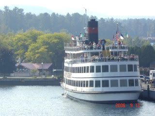
Cruise along on Lake George if you want to drive just a bit further.. lot of other places too.. just quickly spotted these clicks on my hard disk. ;)


But mainly I am going to meet old friends, catch up, check out everybody's latest at the conference, hang out and have some fun!

Hope to post a bit this coming week. Cheerio!

Friday, May 25, 2012

POC Diagnostics - A walk in the PARC

Hey everyone.. I’m back..!

It’s not as if stuff hasn’t been happening around here. Dusty blogs = all work less play. Yesterday I attended a very inspiring talk and I decided today I just have to take an hour off and summarize it here! 

It was Peter Keisel from the Palo Alto Research Center. PARC is an independent subsidiary of Xerox, and consists of about 170 researchers in 4 labs. This work on sophisticated point-of-care opto-fluidic detection from their opto-electronics group was super-exciting! I had been looking forward to meeting Peter Keisel and hearing more about his work. Finally everything came together at the N-Cal OSA meeting yesterday.
  
ref: http://office.microsoft.com/en-us/images/
Diagnostics and testing of water, blood, and other samples often involves counting cells or particles of a certain type, defined by size, spectra or other characteristics. Equipment like flow cytometers are often used for such testing. But their size, cost and complexity limits their availability. Therefore, such testing is often conducted in dedicated labs and testing centers. Depending on the local infrastructure and perishable nature of the test sample, this costly and time consuming effort, sometimes may not even be possible. Hence development of compact, inexpensive and robust devices for point-of-care testing is a critical and active area of research. 

Peter discussed an opto-fluidic detection device developed by his team at PARC. The device contains...